
Echocardiography
|

|
|
What is an Echocardiogram:
An echocardiogram is a test in which ultrasound is used to examine the heart. The equipment is far superior to that used by fishermen. In addition to providing single-dimension images, known as M-mode echo that allows accurate measurement of the heart chambers, the echocardiogram also offers far more sophisticated and advanced imaging.
This is known as two- dimensional (2-D) Echo and is capable of displaying a cross-sectional "slice" of the beating heart, including the chambers, valves and the major blood vessels that exit from the left and right ventricle Echocardiography uses standard two-dimensional, three-dimensional, and Doppler ultrasound to create images of the heart.
Echocardiography has become routinely used in the diagnosis, management, and follow-up of patients with any suspected or known heart diseases. It is one of the most widely used diagnostic tests in cardiology. It can provide a wealth of helpful information, including the size and shape of the heart (internal chamber size quantification), pumping capacity, and the location and extent of any tissue damage. An Echocardiogram can also give physicians other estimates of heart function such as a calculation of the cardiac output, ejection fraction, and diastolic function (how well the heart relaxes).
Echocardiography can help detect cardiomyopathies, such as hypertrophic cardiomyopathy, dilated cardiomyopathy, and many others. The use of Stress Echocardiography may also help determine whether any chest pain or associated symptoms are related to heart disease. The biggest advantage to echocardiography is that it is noninvasive (doesn’t involve breaking the skin or entering body cavities) and has no known risks or side effects.
Not only can an echocardiogram create ultrasound images of heart structures, but it can also produce accurate assessment of the blood flowing through the heart, using pulsed or continuous wave Doppler ultrasound. This allows assessment of both normal and abnormal blood flow through the heart. Color Doppler as well as spectral Doppler is used to visualize any abnormal communications between the left and right side of the heart, any leaking of blood through the valves (valvular regurgitation), and to estimate how well the valves open (or do not open in the case of valvular stenosis).
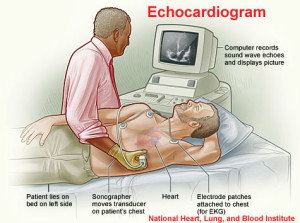
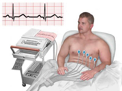
Clothing from the upper body is removed and covered by a gown or sheet to keep you comfortable and maintain the privacy of females. The patient then lies on an examination table or a hospital bed. Sticky patches or electrodes are attached to the chest and shoulders and connected to electrodes or wires. These help to record the electrocardiogram (EKG or ECG) during the echocardiography test. The EKG helps in the timing of various cardiac events (filling and emptying of chambers).
A colorless gel is then applied to the chest and the echo transducer is placed on top of it. The echo technologist then makes recordings from different parts of the chest to obtain several views of the heart. You may be asked to move from your back and to the side. Instructions may also be given for you to breathe slowly or to hold your breath. This helps in obtaining higher quality pictures. The images are constantly viewed on the monitor. It is also recorded on photographic paper and on digital media. The images offer a permanent record of the examination and are reviewed by the physician prior to completion of the final report.
What is a Doppler Examination?
Doppler is a special part of the ultrasound examination that assesses blood flow (direction and velocity). In contrast, the M-mode and 2-D Echo evaluates the size, thickness and movement of heart structures (chambers, valves, etc.). During the Doppler examination, the ultrasound beams will evaluate the flow of blood as it makes its way through and out of the heart. This information is presented visually on the monitor (as color images or grayscale tracings and also as a series of audible signals with a swishing or pulsating sound)..